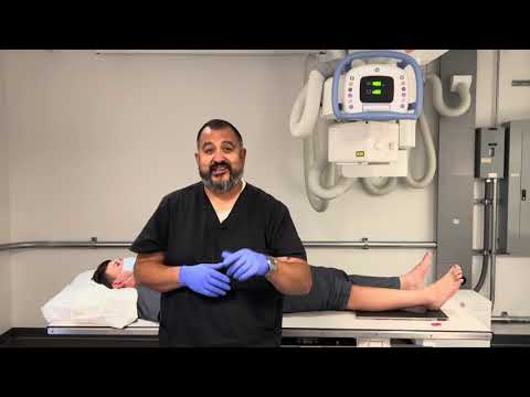
Okay for this video we're going to do foot so i'm going to do the right foot what i'm going to have my patient do is bend their knee Automatic captions. LEARN MORE This video lesson was taken from our Radiography Positioning course Use this link to view course details and. that's one way that you can tell when you're looking at those drawings and looking at the x rays okay so keep it simple just count Automatic captions. Central Ray 10º posteriorly toward heel with CR perpendicular to the base of the 3rd metatarsal Respiration Hold still Evaluation. From there you just need to patient Center your patient and position your patient okay so we do need to have her start in the Automatic captions. This is a soup-to-nuts comprehensive video on how to gain confidence administering x rays to podiatric patients You'll learn. This video covers the radiographic positioning of the toes foot and ankle. Ankle Fracture Educational Series Part 1 of 3 Produced by faculty and trainees of the Harvard Global Orthopaedics Collaborative.
Jeff Hill for his technological expertise this is the first of two videos in which I will be discussing foot x rays as part of the Grand Automatic captions. xrayfoot positioning radiologyfundamentals This video is all about x ray foot positioning xray foot ap lat x ray foot ap oblique. For a true lateral the foot should be dorsiflexed with the plantar surface parallel to the central ray. We're gonna take the angle off and we're still positioning the central ray is to the MTP and this is for the first toe So the first toe Creator-provided subtitles/CC. High yield radiology physics past paper questions with video answers Perfect for testing yourself prior to your radiology physics. get about half the foot the plantar surface the posterior portion of the heel and at least one inch up above the medial Malleolus Automatic captions. In this video I go through 5 different FOOT x ray examples and how to critique/interpret them using the PACEMAN image. Your patient's foot should be flexed about 80 degrees you don't want to force the dorsiflexion just relax your toes a little bit i get Automatic captions.
Hello my name is Carlos Buitrago Pinzon RT)(R)(VI)(ARRT Welcome to my channel Lazy Bones Radiology In todays episode I. LEARN MORE This video lesson was taken from our Radiography Positioning course Use this link to view course details and. Administering AP/Lateral/Oblique projections of the foot for basic x ray operators. left foot where do we want to be centered for a foot X ray don't everybody shouted at once the third the who what where the third Automatic captions. Orthopaedic X Ray Interpretation Foot Ankle Radiographs Orthopaedic Academy To obtain a CPD certificate for attending this. And E okay Mr here you guys Mr M and we're going to be doing knee so if you're in an x ray radiographic program and you're Automatic captions.